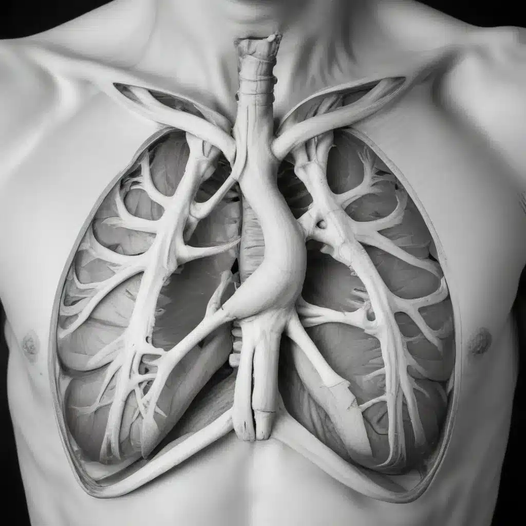
Navigating the complexities of dental care can be a daunting task, but when it comes to the management of rare and potentially life-threatening conditions, it’s crucial for dental professionals to remain vigilant and well-informed. In this comprehensive article, we’ll delve into the intriguing world of pneumomediastinum, an uncommon but ominous condition that can arise as a result of certain dental procedures or trauma. By understanding the underlying mechanisms, recognizing the warning signs, and exploring the latest treatment approaches, we aim to empower both dental practitioners and patients with the knowledge to prevent and effectively manage this invisible menace.
Understanding Pneumomediastinum
Pneumomediastinum is a medical condition characterized by the presence of air or gas in the mediastinum, the central compartment of the thoracic cavity that sits between the lungs. This condition can develop due to a variety of causes, including certain dental procedures, trauma, or underlying medical conditions.
During routine dental treatments, such as extractions, oral surgery, or even endodontic procedures, the delicate balance of air pressure within the body can be disrupted. In some cases, this disruption can allow air to escape from the oral cavity, pharynx, or lungs and enter the mediastinal space, leading to the formation of pneumomediastinum.
The presence of air or gas in the mediastinal area can pose a significant threat to the patient’s health, as it can compress vital structures, such as the trachea, esophagus, and blood vessels. This compression can result in a range of symptoms, including chest pain, difficulty breathing, and even subcutaneous emphysema, a condition where air becomes trapped under the skin.
Recognizing the Warning Signs
Prompt recognition of pneumomediastinum is crucial, as early intervention can greatly improve the patient’s prognosis. Dental professionals should be vigilant in monitoring for the following signs and symptoms during and after dental procedures:
-
Chest Pain: Patients may experience a sharp, stabbing, or burning sensation in the chest, which may radiate to the neck or back.
-
Difficulty Breathing: Patients may report a sensation of tightness in the chest, shortness of breath, or even stridor (a high-pitched, wheezing sound during breathing).
-
Subcutaneous Emphysema: The presence of air trapped under the skin, typically felt as a crackling or crepitus sensation when palpated, can be a telltale sign of pneumomediastinum.
-
Neck Swelling: The accumulation of air in the mediastinal space can cause visible swelling or puffiness in the neck and upper chest area.
-
Voice Changes: Patients may notice changes in their voice, such as hoarseness or a muffled quality, due to the compression of the larynx or trachea.
-
Dysphemia (Difficulty Swallowing): Patients may experience difficulty or discomfort when swallowing due to the compression of the esophagus.
If any of these signs or symptoms are observed during or after a dental procedure, it is crucial for the dental team to act quickly and initiate appropriate diagnostic and treatment measures.
Diagnostic Approach
Prompt and accurate diagnosis is essential in the management of pneumomediastinum. Dental practitioners should have a high index of suspicion and be prepared to coordinate with medical specialists, such as pulmonologists or thoracic surgeons, to ensure a comprehensive evaluation.
The diagnostic workup typically involves the following steps:
-
Physical Examination: A thorough physical examination, including palpation of the neck and upper chest, can reveal the presence of subcutaneous emphysema or other telltale signs of pneumomediastinum.
-
Imaging Studies: Radiographic imaging, such as a chest X-ray or computed tomography (CT) scan, is often the gold standard for confirming the diagnosis of pneumomediastinum. These imaging modalities can help visualize the presence and extent of air or gas in the mediastinal space.
-
Laboratory Tests: In some cases, blood tests may be ordered to evaluate for potential underlying conditions or to assess the patient’s overall health status.
-
Endoscopic Evaluation: In some instances, endoscopic procedures, such as bronchoscopy or esophagoscopy, may be performed to rule out any underlying structural or anatomical abnormalities that may have contributed to the development of pneumomediastinum.
By utilizing a comprehensive diagnostic approach, dental professionals can accurately identify the presence and severity of pneumomediastinum, enabling them to develop an appropriate treatment plan and coordinate care with the necessary medical specialists.
Treatment Strategies
The management of pneumomediastinum requires a multidisciplinary approach, involving close collaboration between dental practitioners, pulmonologists, thoracic surgeons, and other healthcare professionals. The primary goals of treatment are to address the underlying cause, alleviate the patient’s symptoms, and prevent the development of any life-threatening complications.
-
Supportive Care: In mild cases of pneumomediastinum, the initial treatment may involve supplemental oxygen, pain management, and close monitoring of the patient’s vital signs and respiratory status. This approach allows the body to reabsorb the trapped air naturally, as long as there is no significant compromise to the airway or vital structures.
-
Antibiotic Prophylaxis: In cases where the pneumomediastinum is associated with an infection or the presence of an underlying dental condition, the administration of antibiotics may be warranted to prevent the development of any secondary complications.
-
Surgical Intervention: In more severe cases, where the pneumomediastinum is causing significant compression of vital structures or is not resolving with conservative management, surgical intervention may be necessary. This may involve procedures such as mediastinal decompression, chest tube placement, or even surgical exploration to address the underlying cause.
-
Dental Treatment Modifications: For patients who have developed pneumomediastinum as a result of a dental procedure, it is essential to modify the treatment plan and approach to prevent the recurrence of the condition. This may involve the use of alternative techniques, the avoidance of certain procedures, or the implementation of additional precautions to minimize the risk of air or gas introduction into the mediastinal space.
-
Comprehensive Follow-up: Regardless of the initial treatment approach, close monitoring and follow-up care are crucial to ensure the patient’s complete recovery and to identify any potential complications or recurrences of the condition.
By employing a multifaceted treatment strategy and maintaining a collaborative approach with medical specialists, dental professionals can effectively manage pneumomediastinum and minimize the risk of adverse outcomes for their patients.
Real-Life Scenarios and Patient Examples
To illustrate the importance of recognizing and managing pneumomediastinum, let’s consider a few real-life scenarios:
-
Dental Extraction Complication:
Mr. Jones, a 45-year-old patient, presented to a dental clinic for the extraction of a severely impacted wisdom tooth. During the procedure, the dentist noted some difficulty in the extraction and suspected the possibility of a tearing or perforation of the pharyngeal mucosa. Immediately following the extraction, Mr. Jones reported a sudden onset of chest pain and difficulty swallowing. The dental team promptly recognized the signs of pneumomediastinum and coordinated with a thoracic surgeon for further evaluation and management. A CT scan confirmed the presence of air in the mediastinal space, and Mr. Jones underwent a successful mediastinal decompression procedure, followed by a period of observation and antibiotic therapy. He made a full recovery and was able to resume his normal activities within a few weeks. -
Traumatic Pneumomediastinum:
Ms. Garcia, a 32-year-old patient, presented to the dental clinic with facial trauma after a recent car accident. Upon examination, the dentist noted the presence of subcutaneous emphysema in the neck and upper chest area. Suspecting the possibility of pneumomediastinum, the dental team promptly ordered a chest X-ray, which confirmed the diagnosis. Ms. Garcia was immediately referred to the emergency department, where a thoracic surgeon evaluated her condition and determined that the pneumomediastinum was a result of the traumatic injury. She underwent observation and conservative management, which included the administration of supplemental oxygen and pain medication. After a few days of close monitoring, Ms. Garcia’s condition improved, and she was discharged with instructions for follow-up care with her primary care physician.
These real-life scenarios highlight the importance of dental professionals being vigilant in recognizing the signs and symptoms of pneumomediastinum, as well as the necessity of coordinating with medical specialists to ensure prompt and effective management of this potentially life-threatening condition.
The Role of Preventive Measures
While the occurrence of pneumomediastinum in the dental setting is relatively rare, there are proactive steps that dental practitioners can take to minimize the risk and ensure the safety of their patients:
-
Proper Technique and Precautions: Dental professionals should be well-versed in the proper techniques and precautions for procedures that carry a higher risk of air or gas introduction, such as extractions, oral surgery, or endodontic treatments. This may involve the use of specialized equipment, the implementation of suction protocols, and a heightened awareness of the patient’s airway and respiratory status during the procedure.
-
Patient Education and Communication: Educating patients about the potential risks associated with certain dental procedures and empowering them to recognize the warning signs of pneumomediastinum can significantly contribute to early detection and intervention. Dental practitioners should engage in open and transparent communication with their patients, ensuring that they understand the risks and the importance of reporting any concerning symptoms.
-
Comprehensive Emergency Preparedness: Dental clinics should have a well-established emergency response plan in place, which includes the availability of necessary equipment, such as oxygen and suction devices, as well as the training of staff to recognize and respond to respiratory emergencies. Periodic drills and simulations can help ensure that the entire dental team is prepared to handle such situations effectively.
-
Collaboration with Medical Specialists: Maintaining strong relationships and open communication channels with medical specialists, such as pulmonologists, thoracic surgeons, and emergency physicians, can greatly facilitate the seamless coordination of care in the event of a pneumomediastinum diagnosis.
By implementing these preventive measures and fostering a culture of vigilance and preparedness, dental professionals can significantly reduce the risk of pneumomediastinum and ensure the highest level of patient safety.
Embracing Modern Dental Technologies
Advancements in dental technology have not only enhanced the quality of care but have also contributed to the prevention and management of rare conditions like pneumomediastinum. Dental practitioners can leverage the following modern technologies to enhance their diagnostic and treatment capabilities:
-
Digital Imaging: The use of digital radiography and cone-beam computed tomography (CBCT) can provide high-quality, detailed images of the oral and maxillofacial region, enabling early detection of subtle signs of pneumomediastinum or underlying conditions that may predispose a patient to this condition.
-
Endoscopic Procedures: Minimally invasive endoscopic techniques, such as flexible laryngoscopy and esophagoscopy, can be employed to directly visualize the upper airway and esophagus, aiding in the diagnosis and evaluation of any structural abnormalities or the extent of air or gas accumulation.
-
Monitoring and Ventilation Systems: Advanced respiratory monitoring and ventilation equipment can play a crucial role in the management of pneumomediastinum, allowing for the close monitoring of the patient’s respiratory status and the delivery of targeted oxygen therapy or mechanical ventilation, if necessary.
-
Telemedicine and Remote Consultation: In the event of a suspected pneumomediastinum, the ability to remotely consult with medical specialists, such as pulmonologists or thoracic surgeons, can expedite the decision-making process and facilitate the coordination of appropriate care.
By embracing these modern dental technologies, practitioners can enhance their ability to diagnose, monitor, and manage pneumomediastinum, ultimately improving patient outcomes and ensuring the highest standards of dental care.
Conclusion
Pneumomediastinum, though a rare occurrence in the dental setting, is a condition that requires vigilance, prompt recognition, and a multidisciplinary approach to management. By understanding the underlying mechanisms, recognizing the warning signs, and employing a comprehensive diagnostic and treatment strategy, dental professionals can effectively tame this invisible menace and safeguard the well-being of their patients.
Through ongoing education, collaboration with medical specialists, and the strategic implementation of modern dental technologies, the team at Station Road Dental Aldergrove remains committed to providing the highest level of care and ensuring the safety and comfort of our valued patients. Together, we can navigate the complexities of this condition and continue to deliver exceptional dental services that prioritize the overall health and well-being of our community.

